Back to blog
Wellness
X-ray, CT, PET, and MRI: Differences and Uses
4 min read
Ajolote Azul Team
medical imaging
CT scan
MRI
PET scan
radiation dose
genetic testing
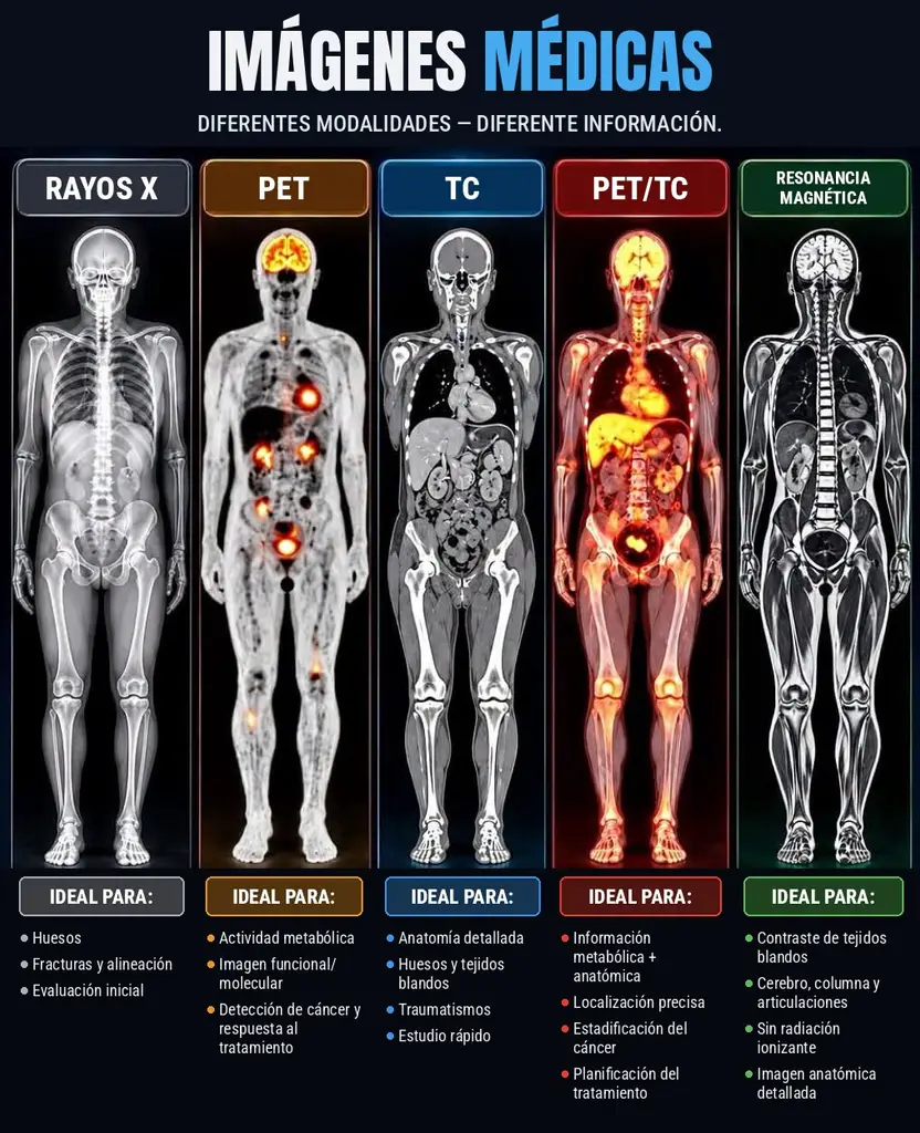
When a doctor orders an imaging study, the choice is not random. Each technique “sees” the body differently: some show structure (anatomy) and others function (metabolism). Here is a simple guide to what each is usually used for.
X-ray: the starting point
A radiograph uses a small dose of ionizing radiation to produce a two-dimensional image. It is quick, widely available, and very useful for bones, fractures, alignment, and as an initial evaluation (for example of the chest). A chest X-ray is roughly 0.1 mSv, similar to about 10 days of natural background radiation.
CT (computed tomography): detailed anatomy
CT takes multiple X-ray images from different angles and a computer combines them into cross-sectional slices. It offers detailed anatomy of bone and soft tissue, and because it is fast it is key in trauma and emergencies. Its radiation dose is higher than a plain X-ray (abdominal and pelvic CT is around 7.7 mSv, according to RadiologyInfo). Contrast is sometimes used, so you may be asked about allergies and kidney function.
PET: metabolism in an image
Positron emission tomography (PET) uses a small amount of radioactive tracer, often a glucose analog (FDG). Cells with higher metabolic activity take up more. PET therefore provides functional and molecular imaging: it shows metabolic activity and is used, among other things, in cancer detection and to assess treatment response. Not every uptake means cancer—inflammation and infections can also light up, and a specialist interprets the scan.
PET/CT: function and anatomy in one session
PET/CT combines both in a single visit: PET shows where activity is and CT shows the exact structure. Linking metabolic and anatomic information allows precise localization and is widely used for cancer staging and treatment planning. It is among the higher-dose studies (about 22.7 mSv for a whole-body protocol per RadiologyInfo), so it is ordered when clinical benefit justifies it.
MRI: soft tissue without ionizing radiation
Magnetic resonance imaging (MRI) uses a strong magnetic field and radio waves—no ionizing radiation. It excels at soft-tissue contrast and detailed anatomy, ideal for brain, spine, and joints. The exam usually takes longer and the machine is noisy and enclosed. Tell the team if you have a pacemaker, implants, prostheses, or metal fragments.
Radiation and preparation: the basics
- Radiation: X-ray, CT, PET, and PET/CT use ionizing radiation; MRI does not. Centers follow the ALARA principle (dose as low as reasonably achievable).
- Pregnancy: always say if you are or could be pregnant.
- PET and PET/CT: several hours of fasting, avoiding intense exercise beforehand, and glucose control in people with diabetes are common instructions.
- CT with contrast: mention allergies, kidney problems, and medications.
- MRI: remove metal objects and mention claustrophobia.
Always follow your imaging center’s specific instructions.
Who chooses which study?
Your physician picks the appropriate test based on your symptoms, history, and the clinical question. More imaging does not always mean a better diagnosis; each study has benefits and risks that must be weighed case by case.
Medical imaging and genetic tests: two complementary views
Imaging shows how your body looks now: structure, lesions, or metabolic activity. Genetic tests show predisposition: inherited variants that may raise risk for certain diseases or influence response to treatments. Together they help your doctor personalize surveillance—for example, someone with hereditary cancer risk may need different follow-up than the general population.
To see which genetic tests we offer and how they can complement medical care, visit us: testgenetico-ajoloteazul.com.
Ajolote Azul Genomics – Exclusive Nutrigenomix distributor in Mexico
This article is informational and does not replace medical advice.
If genetics interests you, you can compare our genetic tests and pricing.
Frequently asked questions
What is the difference between a CT scan and an MRI?
CT uses X-rays, is fast, and is very useful for bone, lung, and emergencies. MRI uses magnets and radio waves, emits no ionizing radiation, and shows soft tissues such as brain, spinal cord, joints, and muscles more clearly.
Does PET involve radiation?
Yes. It uses a small amount of radioactive tracer (often FDG, a form of glucose) that accumulates in tissues with higher metabolic activity. Your doctor weighs benefit against that exposure.
Can imaging replace a genetic test?
No. Imaging shows how your organs are today; a genetic test analyzes DNA variants. They answer different questions and can complement each other.
References
- RadiologyInfo.org (RSNA/ACR). Radiation dose in X-ray and CT exams. https://www.radiologyinfo.org/en/info/safety-xray
- RadiologyInfo.org. Computed tomography (CT) – body. https://www.radiologyinfo.org/en/info/bodyct
- RadiologyInfo.org. PET/CT. https://www.radiologyinfo.org/en/info/pet
- RadiologyInfo.org. Magnetic resonance imaging (MRI) – body. https://www.radiologyinfo.org/en/info/bodymr
- National Institute of Biomedical Imaging and Bioengineering (NIBIB, NIH). Science topics. https://www.nibib.nih.gov/science-education/science-topics
A
Ajolote Azul Team
Specialist in wellness, nutrition and holistic health. Passionate about sharing science-based knowledge to improve quality of life.
Keep Reading
Related Articles
Discover more content on this topic that might interest you
Wellness
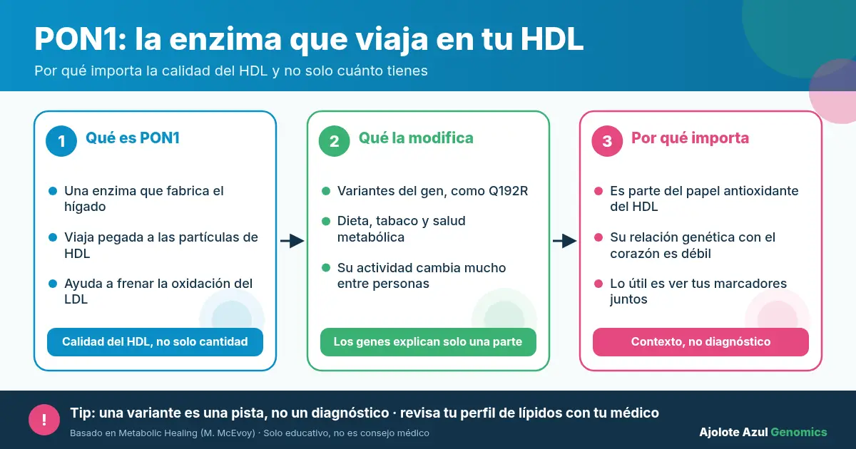
PON1 Gene: Function, HDL, and the Q192R Variant
What does the PON1 gene do? How it relates to paraoxonase, HDL cholesterol, and the Q192R variant—what the evidence shows and its limits.
7 min readRead more
Wellness
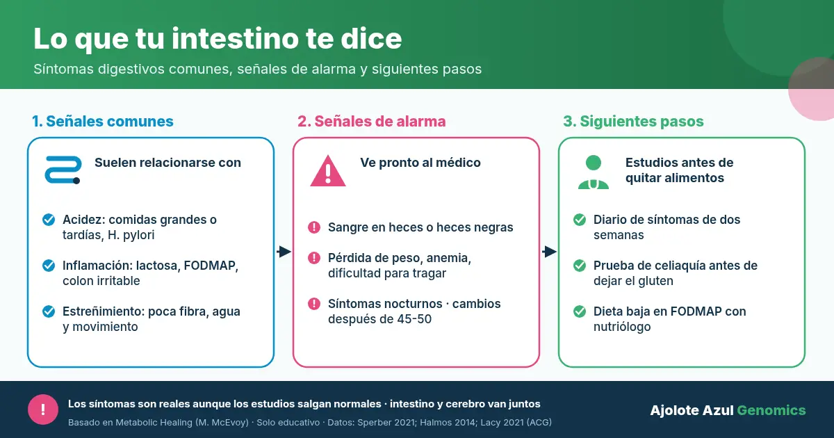
Digestive Symptoms: What Your Gut Is Telling You
Bloating, heartburn, or constipation can have several causes. Learn the warning signs and how a genetic test looks at lactose, gluten, and B12.
8 min readRead more
Wellness
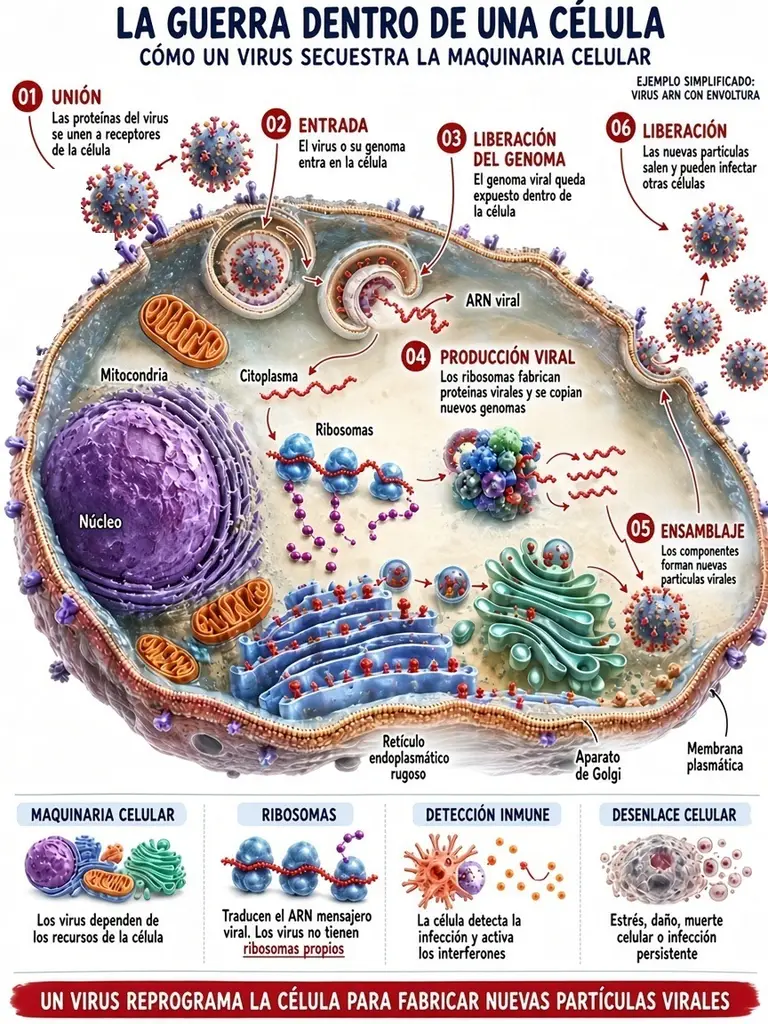
How a Virus Infects a Cell and How the Body Defends
Step by step: how a virus hijacks a cell to multiply, and the defenses your body uses—interferons, sensors, and apoptosis.
5 min readRead more
Did you like this article?
Discover how you can optimize your health with a personalized nutrigenetic test